利用者:空色水色/細胞シグナル伝達
細胞シグナル伝達(米:cell signaling、英:cell signalling)とは、ある細胞が他の細胞に向けて情報伝達物質を放出することである。微小な環境を知覚した上での的確な応答に関与し、発生、組織修復、および免疫の基本を成す。細胞シグナル伝達の異常は癌、自己免疫疾患および糖尿病などの疾患の原因となる[1][2][3] 。細胞シグナル伝達の研究により疾患の治療に役立ち、理論的には、人工組織の作製を可能にする[4]。
システム生物学は細胞シグナル伝達ネットワークの基礎構造および、ネットワークの変化が情報の伝達(シグナル伝達)にどのように影響するかを理解するのに役立つ。細胞シグナル伝達のネットワークは、双安定性や超感度を含む多くの創発現象を示す。ネットワークの解析には、シミュレーションとモデリングおよびその結果解析を含む実験的・理論的アプローチの組み合わせが求められる[5][6] 。長距離アロステリーは多くの場合、細胞シグナル伝達事象の重要な要素である[7]。
細胞間のシグナル伝達
[編集]
細胞シグナル伝達については、単一の生物における細胞間のシグナル伝達ならびにヒト疾患関連について最も研究が進められている。多くの哺乳類において、初期胚細胞は子宮の細胞とシグナルを交換する[8] 。ヒト胃腸管では細菌は他の細菌細胞と、並びに、ヒト上皮細胞および免疫系細胞とシグナルを交換する[9] 。接合に際して出芽酵母Saccharomyces cerevisiaeはペプチドシグナル(接合因子フェロモン)を環境へ分泌して他の細胞に向けて送る。接合因子ペプチドは他の酵母細胞表面の受容体に結合し、接合を準備するよう誘導する[10]。
様式
[編集]細胞シグナル伝達はシグナルの種類によって機械的シグナル伝達と生化学的シグナル伝達がある。機械的シグナルは、細胞に作用する力、もしくは細胞によって生成される力である。これらの力は細胞によって感知され、応答のトリガーとなる[11] 。生化学的シグナルはタンパク質、脂質、イオン、気体などの生化学的分子である。細胞内・細胞間のシグナル伝達は次のように分類される。
- 細胞内分泌 ー シグナルは、産生した細胞を標的とし、シグナル伝達の間はその標的細胞の内部に居続ける。
- 自己分泌 ー シグナルは、産生した細胞を標的にし、細胞外へと分泌され、細胞表面の受容体に作用して標的細胞に影響を及ぼす。白血球などで見られる。
- 接触分泌 - シグナルは、発信細胞と隣接する(接触する)他の細胞を標的とする。このシグナルは、標的の細胞膜のタンパク質成分または脂質成分を介して伝達される。
- 傍分泌 - シグナルは細胞外に分泌されてその近傍の他の細胞を標的とする。シグナルの例は神経伝達物質など。
- 内分泌 - 遠方の細胞を標的とし、血液を介して体内に満遍なく伝達される。内分泌シグナルはホルモンと呼ぶ。一部の細胞のみが特定のホルモンに応答することにより、シグナル伝達が特異性を持つ場合がある。

多くの細胞間コミュニケーションは、直接の細胞間接触を必要とする。多くの細胞は、チャネルにより自身の細胞質を、隣接細胞する細胞質と接続させるギャップ結合ができる。心筋において隣接細胞間のギャップ結合はcardiac pacemaker領域からの活動電位を広げ、心臓の収縮を協調的に引き起こす。
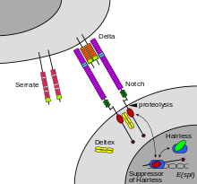
Notchシグナリング機構は接触分泌の様式の一つで、2つの隣接する細胞同士による物理的接触を介したシグナル物質のやり取りである。直接接触が必要条件であることは、胚発生において非常に正確な細胞分化の制御を可能にする。Caenorhabditis elegansにおいて、発生中の生殖腺の2つの細胞は最終分化に至るか、あるいは子宮前駆細胞となり分裂を続ける。 どの細胞が分裂し続けるかは、細胞表面のシグナルの競合によって制御される。どちらかの細胞は、隣接する細胞のNotch受容体を活性化する細胞表面タンパク質をより多く産生する。これは、細胞内のNotch発現を減少させるフィードバックループまたはシステムを活性化させる。[12]
レチノイン酸などの傍分泌シグナルは分泌細胞の近傍の細胞のみを標的とする[13] 。傍分泌シグナル物質には神経伝達物質がある。いくつかのシグナル分子は、ホルモンと神経伝達物質のどちらとしても機能する。例えば、エピネフリンとノルエピネフリンは、副腎から放出されるとホルモンとして機能し、血流によって心臓に運ばれる。ノルエピネフリンはまた、神経細胞によって産生され、脳内の神経伝達物質としても機能する[14] 。エストロゲンは卵巣によって放出され、ホルモンとして機能し、傍分泌または自己分泌シグナル伝達を介して局所的に作用する[15] 。酸素および酸化窒素の活性種は細胞メッセンジャーとしても作用する。このプロセスはredox signalingと呼ばれる。
多細胞生物の細胞シグナル伝達
[編集]多細胞生物において異なる細胞間のシグナル伝達は(短距離への)傍分泌、および(長距離への)内分泌、あるいは接触分泌のいずれかである[16] 。特別な場合として、自己分泌シグナル伝達も起こる[17] 。シナプスシグナル伝達については、化学シナプスの場合はニューロンと標的細胞の間は傍分泌シグナル伝達(化学シナプスの場合)、電気シナプスの場合は接触シグナル伝達である。シグナル分子は、リガンドとして細胞表面受容体へ結合する。次いで/あるいは細胞内分泌シグナル伝達へのエンドサイトーシスによって、または細胞膜を通過して細胞内に侵入することによって標的細胞と相互作用する。これは一般に、セカンドメッセンジャーを活性化させ、様々な生理学的効果をもたらす。
一部のシグナル分子は様々なシグナル伝達様式で使用される。このため、この種のシグナル分子をシグナル伝達様式で分類することはできない。少なくとも3つのシグナル伝達様式による分類が広く認識されているが、その境界は厳密かつ全てのシグナル分子を網羅していない。この伝達様式を示す。
- ホルモンは内分泌の主要なシグナル物質であるが、多くは局所的なシグナル伝達(例えば、ランゲルハンス島細胞)を介す。ほとんどの場合、局所的な目的で発現する(アンジオテンシンなど)。構造的に関連した分子は本来のホルモンによるシグナル伝達を阻害する (PTHrPなど)。
- 神経伝達物質は神経系のシグナル伝達分子であり、神経ペプチドおよび神経調節物質も含む。カテコールアミンのような神経伝達物質も内分泌系によって体循環系に分泌される。
- サイトカインは免疫系の一次傍分泌または接触分泌のシグナル伝達分子であり、鉄代謝か体温の変化を伴う全身作用に関与する。一部の成長因子はサイトカインである。
シグナル分子には様々な種類の化学物質(脂質、リン脂質、アミノ酸、モノアミン、タンパク質、糖タンパク質、各種気体分子)がある。細胞に進入するシグナル分子(グルココルチコイド、甲状腺ホルモン、コレカルシフェロール、レチノイン酸など)は一般的に低分子かつ疎水性で、表面受容体に結合するシグナル分子(TRH、バソプレシン、アセチルコリンなど)は高分子量で親水性である。どちらも例外は多く、同じ分子が表面受容体か内分泌で両方の振舞いを見せるシグナル分子があり、それぞれの振舞いで異なる効果を示す。
硫化水素は人体のいくつかの細胞によって少量生成され、多くのシグナル伝達機能を示す。人体でシグナル分子として作用することが現在知られている他の気体分子には酸化窒素と一酸化炭素がある。[18]
細胞の運動性および分化のための受容体
[編集]細胞は、Notchなどの受容体タンパク質を介して隣接細胞からシグナル分子として情報を受け取る。動物は、Notch受容体と特異的に相互作用することで、細胞表面でのNotchの発現を刺激するシグナルタンパク質をコードする少数の遺伝子セットを持つ。図2に示すように、Notchは、隣接する細胞上に発現するリガンドを刺激する受容体として作用する。Notch受容体相互作用のようなリガンド受容体相互作用は、細胞シグナル伝達のメカニズムおよびコミュニケーションに関与する主な相互作用であることが知られている[19]。
いくつかの受容体は細胞表在性タンパク質であるが、内在性の受容体もある。例えば、エストロゲンは疎水性のシグナル分子であり、脂質二重層を通過し細胞内のエストロゲン受容体を活性化させる。このシグナル伝達は内分泌系の一例であり、エストロゲン受容体は多様な細胞型に見られる。一方、親水性で細胞膜を通過できないシグナル分子は細胞表面受容体に作用する。表在性受容体が受け取ったシグナルは、cAMPなどのセカンドメッセンジャーによって細胞内へと伝達される[20][21]。
シグナル伝達経路
[編集]

いくつかの場合、シグナル分子の受容体活性化はそのシグナル分子に対する細胞応答に直接つながる。例えば、神経伝達物質GABAはイオンチャネルの細胞表面受容体(GABA受容体)を活性化する。GABAが神経細胞のGABAA受容体に結合すると、この受容体の一部である塩化物選択性イオンチャネルが開く。GABAA受容体の活性化は、負に帯電した塩化物イオンがニューロン内に移動することを可能にし、神経細胞の活動電位生成を阻害する。
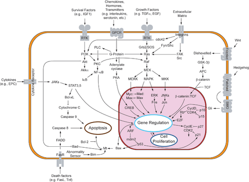
しかし、多くの細胞表面受容体では、リガンド-受容体相互作用は細胞応答に直接関連しない。この場合、活性化受容体は細胞内の他のタンパク質と相互作用する。タンパク質の相互作用は連鎖し、最終的に細胞応答へとつながる。受容体活性化を引き金とする一連の化学反応全体をシグナル伝達機構またはシグナル伝達経路と呼ぶ[22]。
個体間シグナル伝達
[編集]単細胞生物と多細胞生物のどちらでも個体間で分子シグナル伝達が起こり得る。この場合、ある個体がシグナル分子を産生して環境中に分泌・拡散させ、別の個体がシグナル分子を感知または内在化する。発信生物は受信生物の宿主、またはその逆の場合がある。
種間シグナル伝達は特に細菌、酵母、社会性昆虫において起こるが、多くの脊椎動物においても起こる。多細胞生物のシグナル分子はしばしばフェロモンと呼ばれる。種間シグナル伝達には危険を警告する、食糧の存在を教える、繁殖を助けるなどの目的がある.[23]。 単細胞生物ではシグナル伝達は仲間を休眠状態から覚醒させるため、感染力を増大させるため、バクテリオファージに対して防御するためなどに利用される[24]。In quorum sensing, which is also found in social insects, the multiplicity of individual signals has the potentiality to create a positive feedback loop, generating coordinated response. In this context, the signaling molecules are called autoinducers.[25][26][27] This signaling mechanism may have been involved in evolution from unicellular to multicellular organisms.[25][28] Bacteria also use contact-dependent signaling, notably to limit their growth.[29]
Molecular signaling can also occur between individuals of different species. This has been particularly studied in bacteria.[30][31][32] Different bacterial species can coordinate to colonize a host and participate in common quorum sensing.[33] Therapeutic strategies to disrupt this phenomenon are being investigated.[34][35] Interactions mediated through signaling molecules are also thought to occur between the gut flora and their host, as part of their commensal or symbiotic relationship.[35][36] Gram negative microbes deploy bacterial outer membrane vesicles for intra- and inter-species signaling in natural environments and at the host-pathogen interface.
Additionally, interspecies signaling occurs between multicellular organisms. In Vespa mandarinia, individuals release a scent that directs the colony to a food source.[37]
脚注
[編集]- ^ Vlahopoulos, SA; Cen, O; Hengen, N; Agan, J; Moschovi, M; Critselis, E; Adamaki, M; Bacopoulou, F et al. (Aug 2015). “Dynamic aberrant NF-κB spurs tumorigenesis: A new model encompassing the microenvironment”. Cytokine Growth Factor Rev 26: 389–403. doi:10.1016/j.cytogfr.2015.06.001. PMC 4526340. PMID 26119834.
- ^ Wang, K; Grivennikov, SI; Karin, M (2013). “Implications of anti-cytokine therapy in colorectal cancer and autoimmune diseases”. Ann. Rheum. Dis. 72 Suppl 2: ii100–3. doi:10.1136/annrheumdis-2012-202201. PMID 23253923.
- ^ “JNK1 in hematopoietically derived cells contributes to diet-induced inflammation and insulin resistance without affecting obesity”. Cell Metab. 6 (5): 386–97. (2007). doi:10.1016/j.cmet.2007.09.011. PMID 17983584.
- ^ Smith, RJ; Koobatian, MT; Shahini, A; Swartz, DD; Andreadis, ST (2015). “Capture of endothelial cells under flow using immobilized vascular endothelial growth factor”. Biomaterials 51: 303–12. doi:10.1016/j.biomaterials.2015.02.025. PMC 4361797. PMID 25771020.
- ^ Eungdamrong, Narat J.; Iyengar, Ravi (2004-06-01). “Modeling Cell Signaling Networks”. Biology of the Cell 96 (5): 355–362. doi:10.1016/j.biolcel.2004.03.004. ISSN 0248-4900. PMC 3620715. PMID 15207904.
- ^ Dong, P (Sep 2014). “Division of labour between Myc and G1 cyclins in cell cycle commitment and pace control”. Nature Communications 5: 1-11. doi:10.1038/ncomms5750.
- ^ “Proteins MOVE! Protein dynamics and long-range allostery in cell signaling”. Advances in Protein Chemistry and Structural Biology. Advances in Protein Chemistry and Structural Biology 83: 163–221. (2011). doi:10.1016/B978-0-12-381262-9.00005-7. ISBN 978-0-12-381262-9. PMID 21570668.
- ^ “Uterine Wnt/β-catenin signaling is required for implantation”. Proc Natl Acad Sci USA 102 (24): 8579–84. (June 2005). doi:10.1073/pnas.0500612102. PMC 1150820. PMID 15930138.
- ^ “Events at the host-microbial interface of the gastrointestinal tract III. Cell-to-cell signaling among microbial flora, host, and pathogens: there is a whole lot of talking going on”. Am. J. Physiol. Gastrointest. Liver Physiol. 288 (6): G1105–9. (June 2005). doi:10.1152/ajpgi.00572.2004. PMID 15890712.
- ^ “A Microdomain Formed by the Extracellular Ends of the Transmembrane Domains Promotes Activation of the G Protein-Coupled α-Factor Receptor”. Mol Cell Biol 24 (5): 2041–51. (March 2004). doi:10.1128/MCB.24.5.2041-2051.2004. PMC 350546. PMID 14966283.
- ^ Miller, Callie Johnson; Davidson, Lance A. (2013). “The interplay between cell signalling and mechanics in developmental processes”. Nature Reviews Genetics 14 (10): 733–744. doi:10.1038/nrg3513. ISSN 1471-0056. PMC 4056017. PMID 24045690.
- ^ Greenwald I (June 1998). “LIN-12/Notch signaling: lessons from worms and flies”. Genes Dev 12 (12): 1751–62. doi:10.1101/gad.12.12.1751. PMID 9637676.
- ^ Duester G (September 2008). “Retinoic Acid Synthesis and Signaling during Early Organogenesis”. Cell 134 (6): 921–31. doi:10.1016/j.cell.2008.09.002. PMC 2632951. PMID 18805086.
- ^ “Cerebellar norepinephrine modulates learning of delay classical eyeblink conditioning: Evidence for post-synaptic signaling via PKA”. Learn Mem 11 (6): 732–7. (2004). doi:10.1101/lm.83104. PMC 534701. PMID 15537737.
- ^ “Aromatase is abundantly expressed by neonatal rat penis but downregulated in adulthood”. J Mol Endocrinol 33 (2): 343–59. (October 2004). doi:10.1677/jme.1.01548. PMID 15525594.
- ^ Gilbert, Scott F. (2000). “Juxtacrine Signaling”. In NCBI bookshelf. Developmental biology (6. ed.). Sunderland, Mass.: Sinauer Assoc.. ISBN 0878932437
- ^ Bruce Alberts (2002). “General Principles of Cell Communication”. In NCBI bookshelf. Molecular biology of the cell (4th ed.). New York: Garland Science. ISBN 0815332181
- ^ Hausman, Geoffrey M. Cooper, Robert E. (2000). “Signaling Molecules and Their Receptors”. In NCBI bookshelf. The cell : a molecular approach (2nd ed.). Washington, D.C.: ASM Press. ISBN 087893300X
- ^ Cooper, GM (2000年). “The Cell: A Molecular Approach”. www.ncbi.nlm.nih.gov/books/NBK9866/. 2018年9月15日閲覧。
- ^ “Transcriptional Activation by MEIS1A in Response to Protein Kinase A Signaling Requires the Transducers of Regulated CREB Family of CREB Co-activators”. J Biol Chem 284 (28): 18904–12. (2009-07-10). doi:10.1074/jbc.M109.005090. PMC 2707216. PMID 19473990.
- ^ “cAMP-dependent protein kinase A and the dynamics of epithelial cell surface domains: moving membranes to keep in shape”. BioEssays 30 (2): 146–55. (February 2008). doi:10.1002/bies.20705. PMID 18200529.
- ^ Dinasarapu AR (2011). “Signaling gateway molecule pages—a data model perspective”. Bioinformatics 27 (12): 1736–1738. doi:10.1093/bioinformatics/btr190. PMC 3106186. PMID 21505029.
- ^ Tirindelli, R; Dibattista, M; Pifferi, S; Menini, A (July 2009). “From pheromones to behavior”. Physiological Reviews 89 (3): 921–56. doi:10.1152/physrev.00037.2008. PMID 19584317.
- ^ Mukamolova, GV; Kaprelyants, AS; Young, DI; Young, M; Kell, DB (Jul 21, 1998). “A bacterial cytokine”. Proceedings of the National Academy of Sciences of the United States of America 95 (15): 8916–21. doi:10.1073/pnas.95.15.8916. PMC 21177. PMID 9671779.
- ^ a b Miller, Melissa B.; Bassler, Bonnie L. (1 October 2001). “Quorum sensing in bacteria”. Annual Review of Microbiology 55 (1): 165–199. doi:10.1146/annurev.micro.55.1.165. PMID 11544353.
- ^ Kaper, JB; Sperandio, V (June 2005). “Bacterial cell-to-cell signaling in the gastrointestinal tract”. Infection and Immunity 73 (6): 3197–209. doi:10.1128/IAI.73.6.3197-3209.2005. PMC 1111840. PMID 15908344.
- ^ Camilli, A.; Bonnie L. Bassler (24 February 2006). “Bacterial Small-Molecule Signaling Pathways”. Science 311 (5764): 1113–1116. doi:10.1126/science.1121357. PMC 2776824. PMID 16497924.
- ^ Stoka, AM (June 1999). “Phylogeny and evolution of chemical communication: an endocrine approach”. Journal of Molecular Endocrinology 22 (3): 207–25. doi:10.1677/jme.0.0220207. PMID 10343281.
- ^ Blango, Matthew G; Mulvey, Matthew A (31 March 2009). “Bacterial landlines: contact-dependent signaling in bacterial populations”. Current Opinion in Microbiology 12 (2): 177–181. doi:10.1016/j.mib.2009.01.011. PMC 2668724. PMID 19246237.
- ^ Shank, Elizabeth Anne; Kolter, Roberto (31 March 2009). “New developments in microbial interspecies signaling”. Current Opinion in Microbiology 12 (2): 205–214. doi:10.1016/j.mib.2009.01.003. PMC 2709175. PMID 19251475.
- ^ Ryan, R. P.; Dow, J. M. (1 July 2008). “Diffusible signals and interspecies communication in bacteria”. Microbiology 154 (7): 1845–1858. doi:10.1099/mic.0.2008/017871-0.
- ^ Ryan, Robert P.; Dow, J. Maxwell (28 February 2011). “Communication with a growing family: diffusible signal factor (DSF) signaling in bacteria”. Trends in Microbiology 19 (3): 145–152. doi:10.1016/j.tim.2010.12.003. PMID 21227698.
- ^ Déziel, E; Lépine, F; Milot, S; He, J; Mindrinos, MN; Tompkins, RG; Rahme, LG (Feb 3, 2004). “Analysis of Pseudomonas aeruginosa 4-hydroxy-2-alkylquinolines (HAQs) reveals a role for 4-hydroxy-2-heptylquinoline in cell-to-cell communication”. Proceedings of the National Academy of Sciences of the United States of America 101 (5): 1339–44. doi:10.1073/pnas.0307694100. PMC 337054. PMID 14739337.
- ^ Federle, Michael J.; Bassler, Bonnie L. (31 October 2003). “Interspecies communication in bacteria”. Journal of Clinical Investigation 112 (9): 1291–1299. doi:10.1172/JCI20195. PMC 228483. PMID 14597753.
- ^ a b Sperandio, V. (14 July 2003). “Bacteria-host communication: The language of hormones”. Proceedings of the National Academy of Sciences of the United States of America 100 (15): 8951–8956. doi:10.1073/pnas.1537100100. PMC 166419. PMID 12847292.
- ^ Hooper, LV; Gordon, JI (May 11, 2001). “Commensal host-bacterial relationships in the gut”. Science 292 (5519): 1115–1118. doi:10.1126/science.1058709. PMID 11352068.
- ^ Schueller, Teresa I.; Nordheim, Erik V.; Taylor, Benjamin J.; Jeanne, Robert L. (2010). “The cues have it; nest-based, cue-mediated recruitment to carbohydrate resources in a swarm-founding social wasp”. Naturwissenschaften 97 (11): 1017–1022. doi:10.1007/s00114-010-0712-9.
