
利用者:Francesco Nagoya/組織因子


組織因子(そしきいんし、英: tissue factor)は血小板組織因子、第III因子、CD142とも呼ばれるタンパク質であり、血管周囲組織に存在する。[1]チモーゲンであるプロトロンビンからトロンビンを生成する血液凝固カスケードの開始に必要な因子である。歴史的に、血液凝固カスケードは内因系経路と外因系経路が存在すると考えられてきたが、近年では組織因子がカスケードの起点であると考えられるようになっており、外因系経路ではなく組織因子経路と呼ばれることも多い。
機能
[編集]構造
[編集]組織因子は細胞表面に発現する糖タンパク質である。組織因子は第VII因子に対する親和性の高い受容体であり、組織因子/第VII因子複合体は、血液凝固カスケードの起点となっている。血液凝固カスケードを構成する他の因子が不活性な前駆体として血中を循環しているのに対し、組織因子は細胞表面に発現した時点で活性を発揮する。
組織因子は、細胞外ドメイン、膜貫通ドメイン、細胞質ドメインの3つのドメインから成る。組織因子は、先天性の欠損症が報告されていない唯一の血液凝固因子である。[2]
膜型組織因子と同一の遺伝子から、選択的スプライシングにより可溶性組織因子も生成される。可溶性組織因子ではエクソン5が翻訳されておらず、エクソン4とエクソン6が隣接している。[3][4]
凝固
[編集]
組織因子は第VII因子と結合すると、第VII因子の活性化を引き起こし、第VIIa因子-組織因子複合体となる。この複合体はセリンプロテアーゼとして、第IX因子や第X因子を切断して活性化し、組織因子経路と呼ばれる。この作用は、第VIII因子と第IX因子による増幅経路によって増強される。これらの経路により活性化された第X因子は共通経路の起点であり、第V因子やカルシウム、リン脂質の存在下でトロンビンを生成する。この作用は、歴史的にトロンボプラスチン活性と呼ばれてきた。
サイトカイン
[編集]組織因子は、サイトカイン受容体クラスIIファミリーとして知られるタンパク質ファミリーに属する。このファミリーは、その名称が示すとおり、サイトカインと結合することで活性化するものである。第VIIa因子が組織因子に結合すると、細胞内でシグナル伝達が起こり、血管新生やアポトーシスに関係する。[5]
構造
[編集]組織因子は三つのドメインから成る。
- 細胞外ドメインは第VIIa因子と結合する。
- 第VII因子も複数のドメインから成り、そのうちカルボキシル化されたGLA domain (英語)はカルシウムの存在下でリン脂質と結合する。この結合により、組織因子と第VIIa因子の結合は促進される。
- 膜貫通ドメイン
- 細胞内ドメインは21アミノ酸残基からなり、シグナル伝達を担う。
Tissue distribution
[編集]Some cells release TF in response to blood vessel damage (see next paragraph) and some do only in response to inflammatory mediators (endothelial cells/macrophages).
TF is expressed by cells which are normally not exposed to flowing blood such as sub-endothelial cells (e.g. smooth muscle cells) and cells surrounding blood vessels (e.g. fibroblasts). This can change when the blood vessel is damaged by for example physical injury or rupture of atherosclerotic plaques. Exposure of TF expressing cells during injury allows the complex formation of TF with factor VII. Factor VII and TF form an equal molar complex in the presence of calcium ions and this leads to the activation of factor VII on a membrane surface.
The inner surface of the blood vessel consists of endothelial cells. Endothelial cells do not express TF except when they are exposed to inflammatory molecules such as tumor necrosis factor-alpha (TNF-alpha). Another cell type that expresses TF on the cell surface in inflammatory conditions is the monocyte (a white blood cell).
Thromboplastin
[編集]Historically, thromboplastin was a lab reagent, usually derived from placental sources, used to assay prothrombin times (PT time). Thromboplastin, by itself, could activate the extrinsic coagulation pathway. When manipulated in the laboratory, a derivative could be created called partial thromboplastin. Partial thromboplastin was used to measure the intrinsic pathway. This test is called the aPTT, or activated partial thromboplastin time. It was not until much later that the subcomponents of thromboplastin and partial thromboplastin were identified. Thromboplastin is the combination of both phospholipids and tissue factor, both needed in the activation of the extrinsic pathway, and partial thromboplastin is just phospholipids without tissue factor. Tissue factor is not needed to activate the intrinsic pathway.
Interactions
[編集]Tissue factor has been shown to interact with Factor VII.[6][7]
Additional images
[編集]-
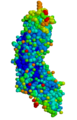 Tissue factor
 -
 Blood plasma after the addition of tissue factor


See also
[編集]References
[編集]- ^ 伊藤正男他編『医学大辞典』第2版、医学書院、2009年、1701頁
- ^ “Entrez Gene: F3 coagulation factor III (thromboplastin, tissue factor)”. 2015年9月21日閲覧。
- ^ “Effect of all-trans retinoic acid and arsenic trioxide on tissue factor expression in acute promyelocytic leukemia cells”. Chin. Med. J. 114 (1): 30–4. (2001). PMID 11779431.
- ^ “Alternatively spliced human tissue factor: a circulating, soluble, thrombogenic protein”. Nat. Med. 9 (4): 458–62. (April 2003). doi:10.1038/nm841. PMID 12652293.
- ^ Marder, Victor J. (2013). Hemostasis and Thrombosis (6th ed.). Philadelphia: Lippincott Williams & Wilkins. pp. 176-177. ISBN 978-1-60831-906-0
- ^ “Probing the interface between factor Xa and tissue factor in the quaternary complex tissue factor-factor VIIa-factor Xa-tissue factor pathway inhibitor”. Eur. J. Biochem. 270 (12): 2576–82. (Jun 2003). doi:10.1046/j.1432-1033.2003.03625.x. PMID 12787023.
- ^ “Structure of extracellular tissue factor complexed with factor VIIa inhibited with a BPTI mutant”. J. Mol. Biol. 285 (5): 2089–104. (Feb 1999). doi:10.1006/jmbi.1998.2452. PMID 9925787.
Further reading
[編集]- “[Initiation in vivo of blood coagulation. The role of white blood cells and tissue factor (author's transl)]”. Nouv Presse Med 8 (40): 3249–53. (1979). PMID 392457.
- “Regulation of the tissue factor gene”. FASEB J. 9 (10): 883–9. (1995). PMID 7615158.
- “Tissue factor pathway”. Baillieres Best Pract. Res. Clin. Haematol. 12 (3): 361–72. (1999). PMID 10856975.
- “The TF:VIIa complex: clinical significance, structure-function relationships and its role in signaling and metastasis”. Thromb. Haemost. 86 (3): 757–71. (2001). PMID 11583305.
- “The pleiotropic effects of tissue factor: a possible role for factor VIIa-induced intracellular signalling?”. Thromb. Haemost. 86 (6): 1353–9. (2001). PMID 11776298.
- “Tissue factor and angiogenesis in cancer”. Curr. Opin. Hematol. 9 (5): 401–6. (2002). doi:10.1097/00062752-200209000-00003. PMID 12172458.
- “The inhibitors of the tissue factor:factor VII pathway”. Thromb. Res. 106 (3): V257-65. (2002). doi:10.1016/S0049-3848(02)00079-8. PMID 12356487.
- “Intravascular tissue factor pathway--a model for rapid initiation of coagulation within the blood vessel”. Thromb. Haemost. 89 (1): 3–8. (2003). doi:10.1267/THRO03010003. PMID 12540946.
- “Tissue factor: in at the start...and the finish?”. J. Thromb. Haemost. 1 (5): 878–80. (2003). doi:10.1046/j.1538-7836.2003.00219.x. PMID 12871349.
- “Oncogenes as regulators of tissue factor expression in cancer: implications for tumor angiogenesis and anti-cancer therapy”. Semin. Thromb. Hemost. 30 (1): 21–30. (2004). doi:10.1055/s-2004-822968. PMID 15034795.
- “Tissue factor and fibrin in tumor angiogenesis”. Semin. Thromb. Hemost. 30 (1): 31–44. (2004). doi:10.1055/s-2004-822969. PMID 15034796.
- “Role of tissue factor in hemostasis, thrombosis, and vascular development”. Arterioscler. Thromb. Vasc. Biol. 24 (6): 1015–22. (2004). doi:10.1161/01.ATV.0000130465.23430.74. PMID 15117736.
- “Signaling of the tissue factor coagulation pathway in angiogenesis and cancer”. Arterioscler. Thromb. Vasc. Biol. 25 (8): 1545–50. (2005). doi:10.1161/01.ATV.0000171155.05809.bf. PMID 15905465.
- “Initiation of coagulation by tissue factor carriers in blood”. Blood Cells Mol. Dis. 36 (2): 188–90. (2007). doi:10.1016/j.bcmd.2005.12.020. PMID 16473535.
- “Cancer-associated thrombosis”. Blood Cells Mol. Dis. 36 (2): 177–81. (2007). doi:10.1016/j.bcmd.2005.12.018. PMID 16490369.
- “Alternatively spliced tissue factor - one cut too many?”. Thromb. Haemost. 97 (1): 5–8. (2007). doi:10.1160/th06-11. PMID 17200762.
- “The changing faces of tissue factor biology. A personal tribute to the understanding of the "extrinsic coagulation activation"”. Thromb. Haemost. 98 (1): 38–42. (2007). doi:10.1160/th07-04-0289. PMID 17597988.
External links
[編集]- Online 'Mendelian Inheritance in Man' (OMIM) 134390
- 蛋白質構造データバンク 今月の分子pdb75_1:日本語分子名(English Name) - March 2006