下腸間膜動脈
表示
| 下腸間膜動脈 | |
|---|---|
 | |
 消化管の腹腔部とその原始・総腸間膜への付着。6週間のヒトの胚(右下に下腸間膜動脈(Inferior mesenteric artery)とある)。 | |
| 概要 | |
| 由来 | 卵黄動脈 |
| 供給源 | 腹大動脈 |
| 分岐 | 左結腸動脈, S状結腸動脈, 上直腸動脈 |
| 静脈 | 下腸間膜静脈 |
| 表記・識別 | |
| ラテン語 | arteria mesenterica inferior |
| MeSH | D017537 |
| グレイ解剖学 | p.609 |
| TA | A12.2.12.069 |
| FMA | 14750 |
| 解剖学用語 | |
下腸間膜動脈(かちょうかんまくどうみゃく、英語: inferior mesenteric artery、しばしばIMAと略される)は、腹大動脈の3番目の主要な分枝である。腰椎3(L3)から生じ、遠位横行結腸から肛門管の上部まで大腸に供給する。IMAが供給する領域は、下行結腸、S状結腸および直腸の一部である[1]。
分布の領域は近接して中結腸動脈と重なり(watershedを形成する)、よって上腸間膜動脈(SMA)と重なる。SMAとIMAは結腸辺縁動脈(Drummond動脈)とリオラン弓(Riolan's arcade, "meandering artery"(蛇行動脈)とも呼ばれ、左結腸動脈と中結腸動脈の間の動脈接続)で吻合する。IMAの分布領域は、胚における後腸とほぼ等しい[1][2]。
分枝
[編集]下腸間膜動脈は、腎動脈の分枝点より下、大動脈分岐部(総腸骨動脈に向かう)より3-4cm上の腰椎3(L3)の高さで腹大動脈の前面から分枝する[3]。
その経路に沿って、IMAには次の分枝がある[1][2][3]。
| 分枝 | 注 |
| 左結腸動脈 | 下行結腸に供給する |
| S状結腸動脈 | 最も上にあり「上S状結腸動脈」と呼ばれる |
| 上直腸動脈 | 事実上、IMAの終枝(他の全ての分枝の後、IMAが続いたもの) |
これら全ての動脈分枝はアーケードにさらに分かれ、一定間隔で結腸に供給される。
関連する静脈
[編集]IMAにはその経路に沿って同じ名前の静脈、下腸間膜静脈が付随しており、これは脾静脈に流れ込む[1]。
よって、下腸間膜静脈は門脈に流れこむため、IMAの経路を完全には反映していない[1][2][3]。
外科と病理
[編集]IMAおよび/またその枝は左半結腸切除術のためには切除する必要がある[4]。
一般的な腎臓の異常(500人に1人)である馬蹄腎は、IMAの下に位置する[5][6]。
図
[編集]-
腹大動脈とその分枝
-
下腸間膜動脈とその分枝
-
腹腔神経叢および下腹神経叢をともなう交感神経幹の腹部
-
上十二指腸陥凹
-
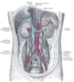
腹膜を取り除いた後の後腹壁。腎臓、腎上体、大血管が示されている。
-
腹部の前面。動脈及び鼠径管の表面マーキングが示されている。
-
下腸間膜動脈
-
腰神経叢と仙骨神経叢 Deep dissection.Anterior view.
-
腰神経叢と仙骨神経叢 Deep dissection.Anterior view.
-
腰神経叢と仙骨神経叢 Deep dissection.Anterior view.
脚注
[編集]- ^ a b c d e Standring, Susan. Gray's anatomy : the anatomical basis of clinical practice (41st ed.). Philadelphia: Elsevier Limited. ISBN 9780702052309. OCLC 920806541
- ^ a b c Moore, Keith L.; Dalley, Arthur F., II; Agur, A. M. R.. Clinically oriented anatomy (7th ed.). Philadelphia: Wolters Kluwer Health/Lippincott Williams & Wilkins. ISBN 1451119453. OCLC 813301028
- ^ a b c Drake, Richard L.; Vogl, Wayne; Mitchell, Adam W. M.; Gray, Henry. Gray's anatomy for students (3rd ed.). Philadelphia, PA: Churchill Livingstone/Elsevier. ISBN 9780702051319. OCLC 881508489
- ^ Charan, Ishwar; Kapoor, Akhil; Singhal, Mukesh Kumar; Jagawat, Namrata; Bhavsar, Deepak; Jain, Vikas; Kumar, Vanita; Kumar, Harvindra Singh (December 2015). “High Ligation of Inferior Mesenteric Artery in Left Colonic and Rectal Cancers: Lymph Node Yield and Survival Benefit”. The Indian Journal of Surgery 77 (Suppl 3): 1103–1108. doi:10.1007/s12262-014-1179-2. ISSN 0972-2068. PMC 4775673. PMID 27011519.
- ^ Schiappacasse, G; Aguirre, J; Soffia, P; Silva, C S; Zilleruelo, N (January 2015). “CT findings of the main pathological conditions associated with horseshoe kidneys”. The British Journal of Radiology 88 (1045). doi:10.1259/bjr.20140456. ISSN 0007-1285. PMC 4277381. PMID 25375751.
- ^ “Clinical case: Horseshoe kidney transplantation”. Kenhub. 2019年9月28日閲覧。
外部リンク
[編集]- Lotti M. Anatomy in relation to left colectomy
- Anatomy figure: 39:02-05 at Human Anatomy Online, SUNY Downstate Medical Center - "Branches of the inferior mesenteric artery."
- Anatomy photo:40:11-0103 at the SUNY Downstate Medical Center - "Posterior Abdominal Wall: Branches of the Abdominal Aorta"
- Anatomy image:7924 at the SUNY Downstate Medical Center
- Anatomy image:7997 at the SUNY Downstate Medical Center
- Anatomy image:8407 at the SUNY Downstate Medical Center
- Anatomy image:8659 at the SUNY Downstate Medical Center
- Atlas image: abdo_wall70 at the University of Michigan Health System - "Posterior Abdominal Wall, Dissection, Anterior View"
- sup&infmesentericart at The Anatomy Lesson by Wesley Norman (Georgetown University)


